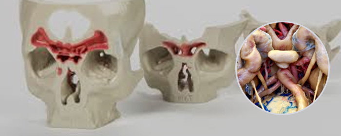


The skull base (or cranial base) is the part of the skull (cranium) that supports the brain and separates the brain from the rest of the head. Blood vessels to the brain and nerves from the brain (cranial nerves) run through holes in the skull base. Below the skull base are the nasal passages, sinus cavities, facial bones, and muscles associated with chewing.
Endoscopic Skull Base Surgery is a form of minimally invasive surgery to correct a number of different conditions affecting the skull base (the region between the brain and back of the nose, mouth and throat). It uses natural body openings (in this case primarily the nose, but sometimes also the mouth) to insert a medical device called an endoscope, which is a thin tube equipped with a camera, lighting, and surgical instruments. Most skull base endoscopic surgery is performed through the nose, in which case it is referred to as an Endoscopic Endonasal Approach (EEA).
Endoscopic Skull Base Surgery may be required to treat any of the following conditions
Benign or malignant tumours (or other abnormal tissue) located underneath the brain, or in the base of the skull or near the uppermost section of the spine, including:
Tumours in / near the pituitary gland (located behind the nose and eyes) e.g. – pituitary adenoma, craniopharyngioma
Tumours in the meninges (a layer of tissue between the brain and skull) – called meningiomas, which are located on the floor of the skull separating the brain and nose
Cysts (Rathke’s cleft cyst)
Abnormal growths caused by infection
Bone tumours at the skull base (chordomas).
Endoscopic skull base surgery is carried out by a multidisciplinary team including a neurosurgeon and an ENT (Ear, Nose and Throat) surgeon - an otolaryngologist. The ENT surgeon makes a small incision inside the nasal cavity which allows the neurosurgeon to operate using the endoscope (described above).
In some cases, the procedure may involve two separate procedures on different days, with the nasal 'exposure' as the first procedure and the tumour removal during the second procedure.
The entire procedure is normally complete within two – four hours, although more complex surgery can take up to six hours to complete. Patients are generally able to get out of bed the day after the procedure.
Most patients are able to go home after several days admission in hospital. Although some may be able to go home the following day, most patients require at least one day in intensive care unit and a few days on the ward for observation and recovery.
Where surgery was via the nasal passage, either nasal packing or a balloon catheter may need to remain in place for up to seven days after surgery. This is then removed, either in hospital or in our offices. Small plastic splints may also need to remain in the nasal passage for up to 14-21 days after surgery. Patients will need to return every 14 days for an endoscopic examination of the nasal cavity to check the healing process, which is generally complete within 3-4 months. Sometimes, an additional drain is inserted in the spine (lumbar drain) to help healing in the nose and preventing leakage of brain fluid.